Copyright
©The Author(s) 2016.
World J Radiol. Sep 28, 2016; 8(9): 764-774
Published online Sep 28, 2016. doi: 10.4329/wjr.v8.i9.764
Published online Sep 28, 2016. doi: 10.4329/wjr.v8.i9.764
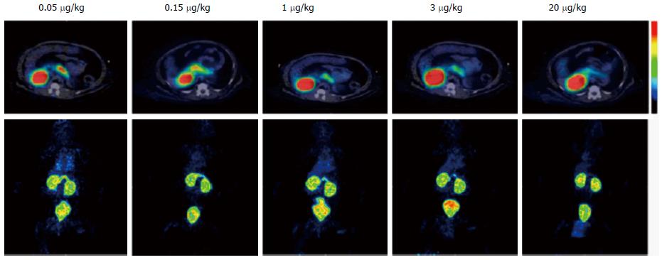
Figure 5 Positron emission tomography/computed tomography images of 68Ga-DO3A-exendin-4 for cynomolgus monkeys.
Increasing concentration of unlabeled peptide resulted in competition for glucagon-like peptide-1 receptor in pancreas only. Transaxial images (dynamic sequences 30-90 min, top row) and whole-body maximum-intensity projections (dynamic sequences 90-120 min, bottom row). Reprinted with permission from Ref. [27].
- Citation: Li J, Karunananthan J, Pelham B, Kandeel F. Imaging pancreatic islet cells by positron emission tomography. World J Radiol 2016; 8(9): 764-774
- URL: https://www.wjgnet.com/1949-8470/full/v8/i9/764.htm
- DOI: https://dx.doi.org/10.4329/wjr.v8.i9.764


