Copyright
©The Author(s) 2016.
World J Radiol. Aug 28, 2016; 8(8): 743-749
Published online Aug 28, 2016. doi: 10.4329/wjr.v8.i8.743
Published online Aug 28, 2016. doi: 10.4329/wjr.v8.i8.743
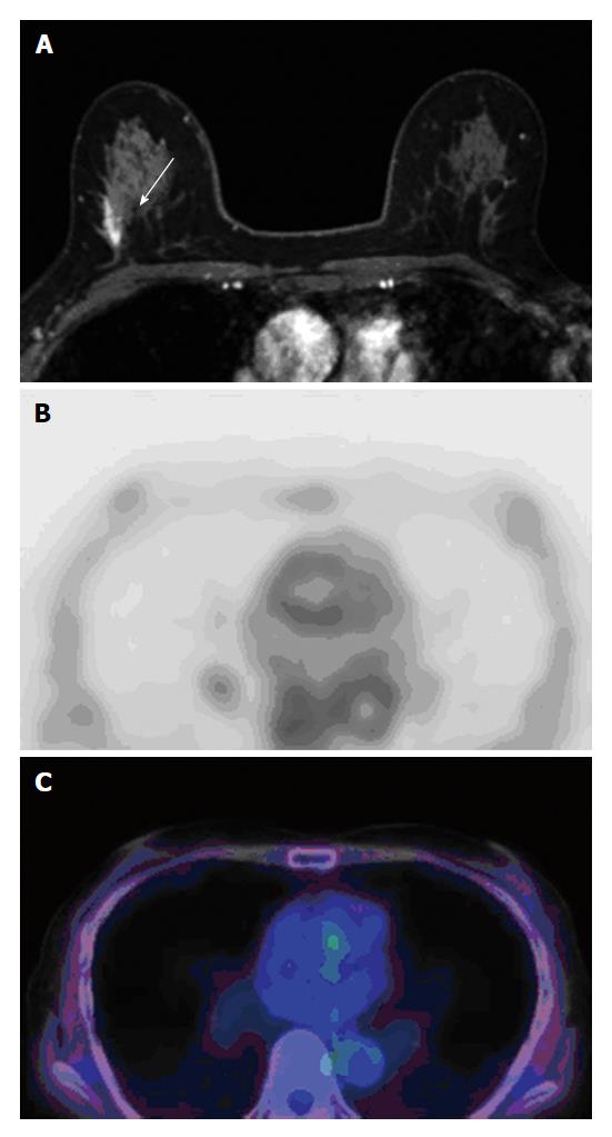
Figure 2 A 64-year-old woman with mammographically detected microcalcifications in right breast was diagnosed with ductal carcinoma in situ using stereotactic stereotactic vacuum-assisted biopsy.
MRI shows a 12-mm non mass enhancement in the right breast (A, arrow). [F-18] FDG PET/CT did not depict any abnormal uptake (B and C). On histopathologic examination, a 15-mm DCIS (absence of comedo necrosis, ER positive, PgR positive, HER-2 positive, nuclear grade1 DCIS) was found. MRI: Magnetic resonance imaging; FDG PET/CT: Fluorodeoxyglucose-positron emission tomography/computed tomography; DCIS: Ductal carcinoma in situ; ER: Estrogen receptor; PgR: Progesterone receptor; HER: Human epidermal growth factor receptor.
- Citation: Fujioka T, Kubota K, Toriihara A, Machida Y, Okazawa K, Nakagawa T, Saida Y, Tateishi U. Tumor characteristics of ductal carcinoma in situ of breast visualized on [F-18] fluorodeoxyglucose-positron emission tomography/computed tomography: Results from a retrospective study. World J Radiol 2016; 8(8): 743-749
- URL: https://www.wjgnet.com/1949-8470/full/v8/i8/743.htm
- DOI: https://dx.doi.org/10.4329/wjr.v8.i8.743


