Copyright
©The Author(s) 2016.
World J Radiol. Aug 28, 2016; 8(8): 729-734
Published online Aug 28, 2016. doi: 10.4329/wjr.v8.i8.729
Published online Aug 28, 2016. doi: 10.4329/wjr.v8.i8.729
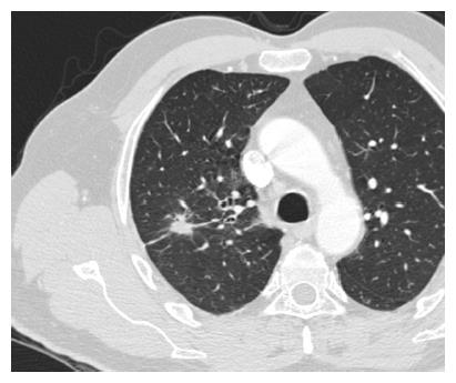
Figure 6 Typical appearance of a malignant solid pulmonary nodule at high-resolution computed tomography (Philips icomputed tomography, slice thickness 1.
25 mm) in a 59-year-old male patient. The lesion was located in the right upper lobe, had a maximum diameter of 24 mm and proved to be an adenocarcinoma at surgery.
- Citation: Perandini S, Soardi GA, Motton M, Augelli R, Dallaserra C, Puntel G, Rossi A, Sala G, Signorini M, Spezia L, Zamboni F, Montemezzi S. Enhanced characterization of solid solitary pulmonary nodules with Bayesian analysis-based computer-aided diagnosis. World J Radiol 2016; 8(8): 729-734
- URL: https://www.wjgnet.com/1949-8470/full/v8/i8/729.htm
- DOI: https://dx.doi.org/10.4329/wjr.v8.i8.729


