Copyright
©The Author(s) 2016.
World J Radiol. Jul 28, 2016; 8(7): 716-725
Published online Jul 28, 2016. doi: 10.4329/wjr.v8.i7.716
Published online Jul 28, 2016. doi: 10.4329/wjr.v8.i7.716
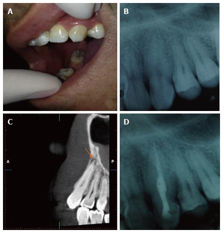
Figure 5 Clinical and radiological assessment of maxillary canine tooth.
A: Clinical appearance of the canine tooth; B: Periapical radiographic findings showed no periapical pathology or carious lesion; C: Sagittal CBCT images showed a periapical radiolucency; D: Periapcial radiography of the treated tooth. The same periapical and CBCT units and settings with Case 1, 2, 3 and 4 were used. CBCT: Cone beam computed tomography.
- Citation: Yılmaz F, Kamburoglu K, Yeta NY, Öztan MD. Cone beam computed tomography aided diagnosis and treatment of endodontic cases: Critical analysis. World J Radiol 2016; 8(7): 716-725
- URL: https://www.wjgnet.com/1949-8470/full/v8/i7/716.htm
- DOI: https://dx.doi.org/10.4329/wjr.v8.i7.716


