Copyright
©The Author(s) 2016.
World J Radiol. Jul 28, 2016; 8(7): 707-715
Published online Jul 28, 2016. doi: 10.4329/wjr.v8.i7.707
Published online Jul 28, 2016. doi: 10.4329/wjr.v8.i7.707
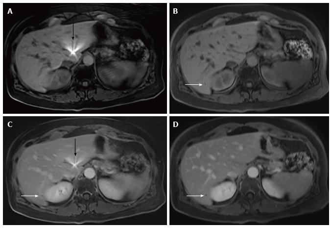
Figure 4 Magnetic resonance imaging of the abdomen in a 53-year-old female with breast cancer performed to follow-up of a pancreatic cyst.
Axial pre-contrast rVIBE (A), pre-contrast cVIBE (B), post-contrast rVIBE (C), and post-contrast cVIBE (D) show superior lesion conspicuity of a tiny hepatic cyst in segment 7 of the liver (white arrows) on pre- and post-contrast cVIBE compared with post-contrast rVIBE images. The lesion was not identified on the pre-contrast rVIBE image. The pre- and post-contrast rVIBE images also illustrate a smooth radiating artifact (black arrow) in the left liver. VIBE: Volumetric interpolated breath-hold examination; rVIBE: Radial VIBE; cVIBE: Cartesian VIBE.
- Citation: Yedururi S, Kang HC, Wei W, Wagner-Bartak NA, Marcal LP, Stafford RJ, Willis BJ, Szklaruk J. Free-breathing radial volumetric interpolated breath-hold examination vs breath-hold cartesian volumetric interpolated breath-hold examination magnetic resonance imaging of the liver at 1.5T. World J Radiol 2016; 8(7): 707-715
- URL: https://www.wjgnet.com/1949-8470/full/v8/i7/707.htm
- DOI: https://dx.doi.org/10.4329/wjr.v8.i7.707


