Copyright
©The Author(s) 2016.
World J Radiol. Jul 28, 2016; 8(7): 700-706
Published online Jul 28, 2016. doi: 10.4329/wjr.v8.i7.700
Published online Jul 28, 2016. doi: 10.4329/wjr.v8.i7.700
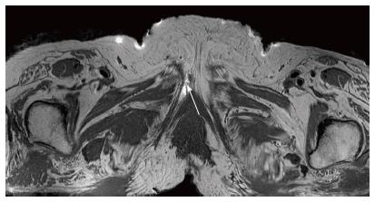
Figure 6 Female pelvis block-post marking.
Identification of marker (arrow) placed in female pelvis block at distal end of inferior pubic ramus canal around dorsal nerve to the clitoris. Contralateral nerve is not well seen.
- Citation: Chhabra A, McKenna CA, Wadhwa V, Thawait GK, Carrino JA, Lees GP, Dellon AL. 3T magnetic resonance neurography of pudendal nerve with cadaveric dissection correlation. World J Radiol 2016; 8(7): 700-706
- URL: https://www.wjgnet.com/1949-8470/full/v8/i7/700.htm
- DOI: https://dx.doi.org/10.4329/wjr.v8.i7.700


