Copyright
©The Author(s) 2016.
World J Radiol. Jul 28, 2016; 8(7): 700-706
Published online Jul 28, 2016. doi: 10.4329/wjr.v8.i7.700
Published online Jul 28, 2016. doi: 10.4329/wjr.v8.i7.700
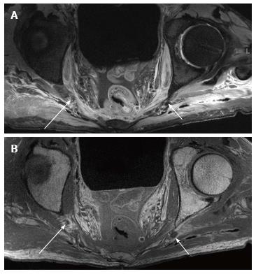
Figure 3 Female pelvic block- premarking.
Axial T2W SPAIR (A) and T1W (B) images show the right (large arrows) and left (small arrows) pudendal nerves at the ischial spine.
- Citation: Chhabra A, McKenna CA, Wadhwa V, Thawait GK, Carrino JA, Lees GP, Dellon AL. 3T magnetic resonance neurography of pudendal nerve with cadaveric dissection correlation. World J Radiol 2016; 8(7): 700-706
- URL: https://www.wjgnet.com/1949-8470/full/v8/i7/700.htm
- DOI: https://dx.doi.org/10.4329/wjr.v8.i7.700


