Copyright
©The Author(s) 2016.
World J Radiol. Jul 28, 2016; 8(7): 668-682
Published online Jul 28, 2016. doi: 10.4329/wjr.v8.i7.668
Published online Jul 28, 2016. doi: 10.4329/wjr.v8.i7.668
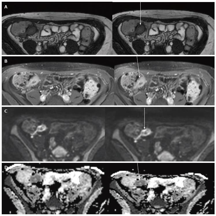
Figure 8 Magnetic resonance enterography of 16-year-old with Crohn’s disease.
Axial true-FISP (4.3/2.2, 50° flip angle) A and contrast axial T1 VIBE B show marked thickening with evidence of enhancement of the wall of terminal ileum, that shows hypersignal on diffusion weighted Imaging (b = 800) image C and restriction of diffusion on ADC map D (arrows).
- Citation: Masselli G, Mastroiacovo I, De Marco E, Francione G, Casciani E, Polettini E, Gualdi G. Current tecniques and new perpectives research of magnetic resonance enterography in pediatric Crohn’s disease. World J Radiol 2016; 8(7): 668-682
- URL: https://www.wjgnet.com/1949-8470/full/v8/i7/668.htm
- DOI: https://dx.doi.org/10.4329/wjr.v8.i7.668


