Copyright
©The Author(s) 2016.
World J Radiol. Jul 28, 2016; 8(7): 656-667
Published online Jul 28, 2016. doi: 10.4329/wjr.v8.i7.656
Published online Jul 28, 2016. doi: 10.4329/wjr.v8.i7.656
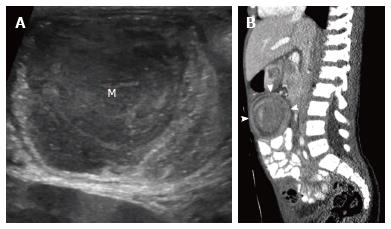
Figure 5 Burkitt lymphoma.
Eight-year-old boy presented with weight loss and abdominal pain. Abdominal ultrasound showed ileocolic intussusception with soft tissue mass (M) as a lead point (A). Bilateral renal masses were also present (not shown). Sagittal reformatted computed tomography image shows ileocolic intussusception (B, arrowheads). Diagnosis was confirmed with biopsy.
- Citation: Gale HI, Gee MS, Westra SJ, Nimkin K. Abdominal ultrasonography of the pediatric gastrointestinal tract. World J Radiol 2016; 8(7): 656-667
- URL: https://www.wjgnet.com/1949-8470/full/v8/i7/656.htm
- DOI: https://dx.doi.org/10.4329/wjr.v8.i7.656


