Copyright
©The Author(s) 2016.
World J Radiol. Jul 28, 2016; 8(7): 656-667
Published online Jul 28, 2016. doi: 10.4329/wjr.v8.i7.656
Published online Jul 28, 2016. doi: 10.4329/wjr.v8.i7.656
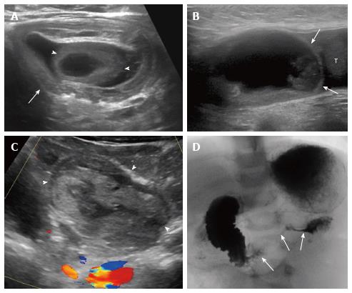
Figure 3 Acquired bowel disorders.
A: Ileocolic intussusception with Meckel diverticulum as lead point. Six-month-old with small bowel obstruction on radiograph (not shown) and intussusception (arrow) demonstrated on ultrasound with lead point (arrowheads); B: Incarcerated inguinal hernia. Two-year-old boy with abdominal pain and left groin mass. Sagittal image of the left inguinal region show a cystic structure that did not clearly communicate with abdominal bowel loops (B, arrows; T = testicle). Testicular edema was also noted (not shown); C, D: Duodenal hematoma secondary to child abuse. One-year-old with abdominal pain and distension. Sagittal midline ultrasound image shows a complex mass in the expected location of the duodenum (C, arrowheads). Upper gastrointestinal series confirmed duodenal narrowing (D, arrows). Abuse was later confirmed.
- Citation: Gale HI, Gee MS, Westra SJ, Nimkin K. Abdominal ultrasonography of the pediatric gastrointestinal tract. World J Radiol 2016; 8(7): 656-667
- URL: https://www.wjgnet.com/1949-8470/full/v8/i7/656.htm
- DOI: https://dx.doi.org/10.4329/wjr.v8.i7.656


