Copyright
©The Author(s) 2016.
World J Radiol. Jul 28, 2016; 8(7): 656-667
Published online Jul 28, 2016. doi: 10.4329/wjr.v8.i7.656
Published online Jul 28, 2016. doi: 10.4329/wjr.v8.i7.656
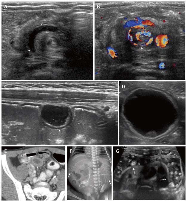
Figure 2 Congenital bowel abnormalities.
A, B: Malrotation. Unsuspected finding in an 8-wk-old vomiting infant being evaluated for pyloric stenosis. Transverse midline images show alternating rings of high and low echogenicity with “whirlpool” sign on grayscale (A) and color Doppler (B) images (arrowheads); C: Gastric duplication cyst. Five-year-old girl with enteric duplication cyst near the gastroesophageal junction detected prenatally (not shown). A second cyst was noted incidentally in the anterior wall of the stomach on subsequent imaging. The cyst demonstrates bowel signature (2 layers), and shares its hypoechoic, muscularis propria layer with the anterior gastric wall (arrowheads); D, E: Meckel diverticulum. Four-year-old with abdominal pain. Ultrasound shows a cyst with bowel signature (D). Computed tomography abdomen is shown for correlation (E, arrow); F, G: Rectourinary fistula with enteroliths. Newborn with abdominal calcifications on radiograph (F, arrows); confirmed to be enteroliths on ultrasound (G, arrows). The fistula was later confirmed with contrast enema (not shown).
- Citation: Gale HI, Gee MS, Westra SJ, Nimkin K. Abdominal ultrasonography of the pediatric gastrointestinal tract. World J Radiol 2016; 8(7): 656-667
- URL: https://www.wjgnet.com/1949-8470/full/v8/i7/656.htm
- DOI: https://dx.doi.org/10.4329/wjr.v8.i7.656


