Copyright
©The Author(s) 2016.
World J Radiol. Jun 28, 2016; 8(6): 635-655
Published online Jun 28, 2016. doi: 10.4329/wjr.v8.i6.635
Published online Jun 28, 2016. doi: 10.4329/wjr.v8.i6.635
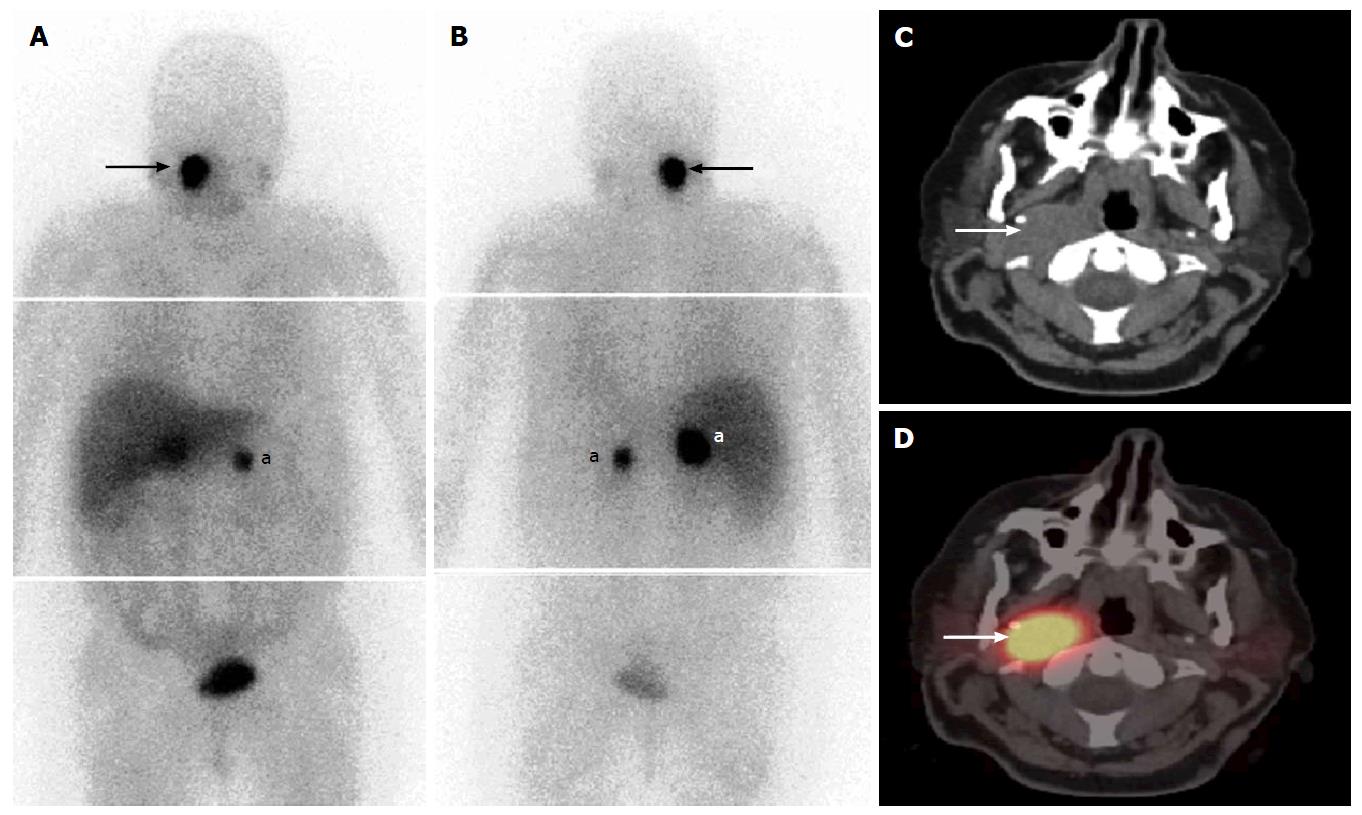
Figure 6 Fifty-three-year-old woman with uncontrolled hypertension and catecholamine hypersecretion, suspected for pheochromocytoma[123].
I-MIBG composite (A) anterior and (B) posterior whole-body images at 24 h post-injection show bilateral focal intense activity in the region of the adrenal glands (a), with unexpected MIBG avid mass in the right side of the neck (arrows). Axial fused SPECT/CT and CT (C, D) images clearly depict a right neck soft tissue mass with intense MIBG uptake (arrows). This indicates the presence of an extra-adrenal head and neck paraganglioma. SPECT: Single photon emission computed tomography; CT: Computed tomography; MIBG: Metaiodobenzylguanidine.
- Citation: Wong KK, Gandhi A, Viglianti BL, Fig LM, Rubello D, Gross MD. Endocrine radionuclide scintigraphy with fusion single photon emission computed tomography/computed tomography. World J Radiol 2016; 8(6): 635-655
- URL: https://www.wjgnet.com/1949-8470/full/v8/i6/635.htm
- DOI: https://dx.doi.org/10.4329/wjr.v8.i6.635


