Copyright
©The Author(s) 2016.
World J Radiol. Jun 28, 2016; 8(6): 635-655
Published online Jun 28, 2016. doi: 10.4329/wjr.v8.i6.635
Published online Jun 28, 2016. doi: 10.4329/wjr.v8.i6.635
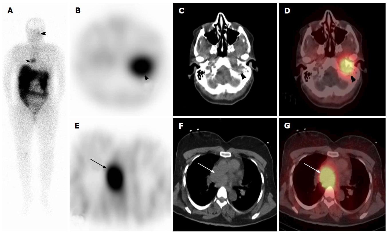
Figure 4 111In-DTPA-pentetreotide scan in a 45-year-old woman with paraganglioma and glomus tumor, to evaluate for octreotide-avidity.
On anterior (A) whole body planar images, there are 2 foci of uptake overlying the left skull base (arrowhead) and the mediastinum (arrows). On Axial SPECT, CT and SPECT/CT (B-D) the skull base radiotracer uptake localizes to a known glomus jugulare tumor adjacent to the left temporal bone and on thoracic axial SPECT, CT and SPECT/CT (E-G) the thoracic radioactivity is within a soft tissue mass in the middle mediastinum, located posterior to the ascending aorta. There is no abnormal radiotracer accumulation below the diaphragm. SPECT: Single photon emission computed tomography; CT: Computed tomography.
- Citation: Wong KK, Gandhi A, Viglianti BL, Fig LM, Rubello D, Gross MD. Endocrine radionuclide scintigraphy with fusion single photon emission computed tomography/computed tomography. World J Radiol 2016; 8(6): 635-655
- URL: https://www.wjgnet.com/1949-8470/full/v8/i6/635.htm
- DOI: https://dx.doi.org/10.4329/wjr.v8.i6.635


