Copyright
©The Author(s) 2016.
World J Radiol. Jun 28, 2016; 8(6): 635-655
Published online Jun 28, 2016. doi: 10.4329/wjr.v8.i6.635
Published online Jun 28, 2016. doi: 10.4329/wjr.v8.i6.635
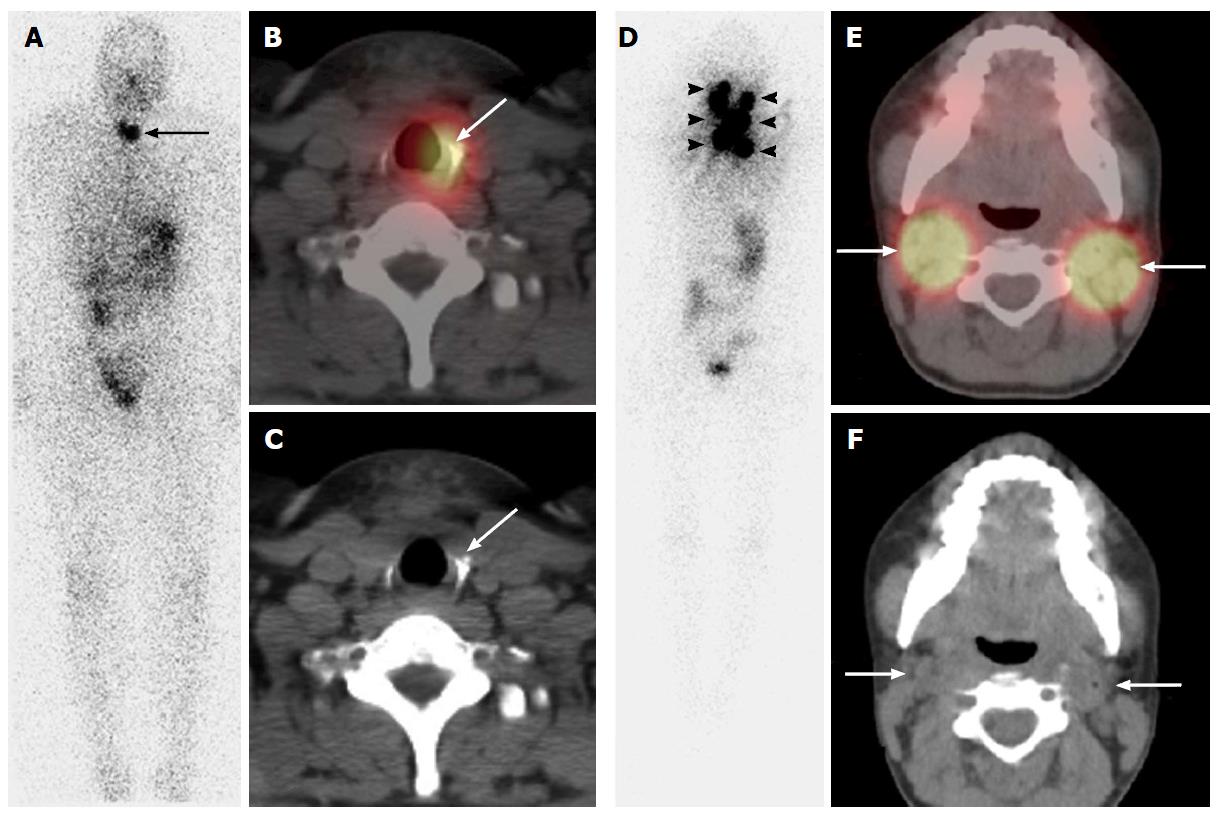
Figure 2 Pre-ablation 131I single photon emission computed tomography/computed tomography scan in a 29-year-old woman with papillary thyroid cancer, the largest lesion measuring 4.
2 cm in the right lobe. Post-surgical thyroglobulin was 0.9 ng/mL with a thyrotropin (TSH) of 57 mIU/L. Planar scan (A) depicts two foci of neck activity (long arrow). SPECT/CT (B, C) localized uptake to soft tissue in the left thyroid bed compatible with remnant thyroid tissue (long arrows) and the patient was given a low dose 131I treatment for remnant ablation. Pre-ablation 131I SPECT/CT scan in a 18-year-old woman with multifocal papillary thyroid cancer, the largest lesion measuring 2.0 cm in the left lobe, with capsular invasion;metastatic lymph nodes were resected from the central compartment. Post-surgical thyroglobulin was 223 ng/mL with a TSH of 68 mIU/L. Planar scan (D) depicts multiple foci of neck activity (arrowheads). SPECT/CT (E,F) localized uptake to bilateral bulky iodine-avid lymph nodes in the neck (long arrows), and also the left supraclavicular fossae (not shown). After imaging the patient classified as high risk for recurrence, recommendation was for medium dose 131I therapy. SPECT: Single photon emission computed tomography; CT: Computed tomography.
- Citation: Wong KK, Gandhi A, Viglianti BL, Fig LM, Rubello D, Gross MD. Endocrine radionuclide scintigraphy with fusion single photon emission computed tomography/computed tomography. World J Radiol 2016; 8(6): 635-655
- URL: https://www.wjgnet.com/1949-8470/full/v8/i6/635.htm
- DOI: https://dx.doi.org/10.4329/wjr.v8.i6.635


