Copyright
©The Author(s) 2016.
World J Radiol. Jun 28, 2016; 8(6): 635-655
Published online Jun 28, 2016. doi: 10.4329/wjr.v8.i6.635
Published online Jun 28, 2016. doi: 10.4329/wjr.v8.i6.635
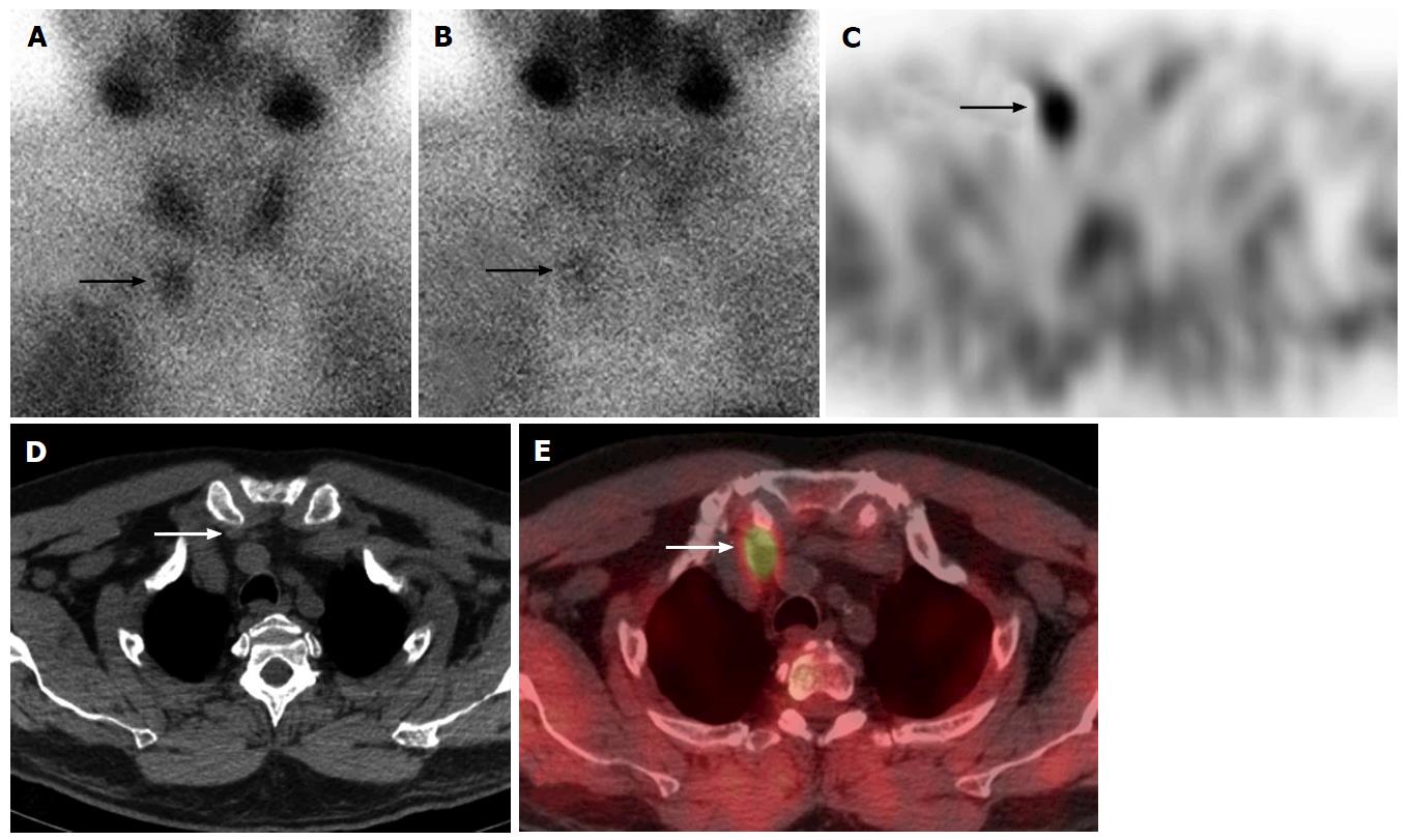
Figure 1 Right inferior parathyroid adenoma in a 60-year-old man.
99mTc-sestamibi parathyroid scan anterior early 20 min (A) and delayed 2 h planar images (B) show focal radiotracer uptake and retention at the juncton of the right lower neck/superior mediastinum (arrows). Axial SPECT, CT and SPECT/CT (C-E) imaging localizes this activity to a soft tissue abnormality in the right lower neck posterior to the sternoclavicular joint (arrows), consistent with ectopic inferior parathyroid adenoma in the lower neck. This was surgically removed via a lower neck incision approach. SPECT: Single photon emission computed tomography; CT: Computed tomography.
- Citation: Wong KK, Gandhi A, Viglianti BL, Fig LM, Rubello D, Gross MD. Endocrine radionuclide scintigraphy with fusion single photon emission computed tomography/computed tomography. World J Radiol 2016; 8(6): 635-655
- URL: https://www.wjgnet.com/1949-8470/full/v8/i6/635.htm
- DOI: https://dx.doi.org/10.4329/wjr.v8.i6.635


