Copyright
©The Author(s) 2016.
World J Radiol. Jun 28, 2016; 8(6): 581-587
Published online Jun 28, 2016. doi: 10.4329/wjr.v8.i6.581
Published online Jun 28, 2016. doi: 10.4329/wjr.v8.i6.581
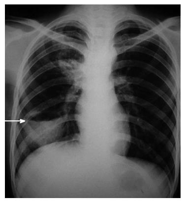
Figure 3 Complicated hydatid cyst: Chest radiograph of a patient who was a known case of hydatid cyst and presented with fever.
Air fluid level can be seen in the cyst (arrow) suggesting superimposed infection. Pyogenic lung abscess is the most common differential diagnosis for this radiological picture and computed tomography may be required to establish the diagnosis.
- Citation: Garg MK, Sharma M, Gulati A, Gorsi U, Aggarwal AN, Agarwal R, Khandelwal N. Imaging in pulmonary hydatid cysts. World J Radiol 2016; 8(6): 581-587
- URL: https://www.wjgnet.com/1949-8470/full/v8/i6/581.htm
- DOI: https://dx.doi.org/10.4329/wjr.v8.i6.581


