Copyright
©The Author(s) 2016.
World J Radiol. Jun 28, 2016; 8(6): 571-580
Published online Jun 28, 2016. doi: 10.4329/wjr.v8.i6.571
Published online Jun 28, 2016. doi: 10.4329/wjr.v8.i6.571
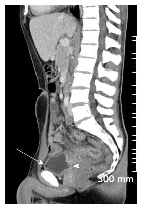
Figure 18 Sagittal oral and intravenous contrast enhanced computed tomography image demonstrating fistulating Crohn’s disease.
There is a wide enterovesical fistula extending from inflamed small bowel to the posterior wall of the bladder (white arrowhead). There is a pocket of air in the bladder (white arrow), indicative of communication with bowel.
- Citation: Stanley E, Moriarty HK, Cronin CG. Advanced multimodality imaging of inflammatory bowel disease in 2015: An update. World J Radiol 2016; 8(6): 571-580
- URL: https://www.wjgnet.com/1949-8470/full/v8/i6/571.htm
- DOI: https://dx.doi.org/10.4329/wjr.v8.i6.571


