Copyright
©The Author(s) 2016.
World J Radiol. Jun 28, 2016; 8(6): 571-580
Published online Jun 28, 2016. doi: 10.4329/wjr.v8.i6.571
Published online Jun 28, 2016. doi: 10.4329/wjr.v8.i6.571
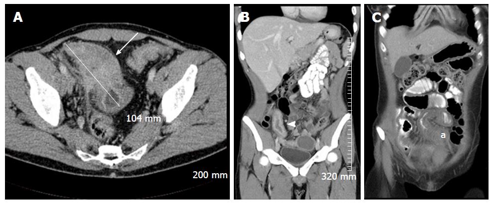
Figure 13 Selected intravenous and oral contrast enhanced computed tomography images of three different patients with acute Crohn’s disease, demonstrating diffuse small bowel mural thickening and enhancement.
A: An axial image demonstrating marked inflammatory thickening of the terminal ileum over approximately 10.4 cm, with extensive adjacent mesenteric stranding (white arrow); B: A coronal image demonstrating marked mural enhancement of a thickened terminal ileum, secondary to terminal ileitis (white arrowhead); C: A coronal image demonstrating inflammatory mural thickening of the mid ileum (a).
- Citation: Stanley E, Moriarty HK, Cronin CG. Advanced multimodality imaging of inflammatory bowel disease in 2015: An update. World J Radiol 2016; 8(6): 571-580
- URL: https://www.wjgnet.com/1949-8470/full/v8/i6/571.htm
- DOI: https://dx.doi.org/10.4329/wjr.v8.i6.571


