Copyright
©The Author(s) 2016.
World J Radiol. Jun 28, 2016; 8(6): 571-580
Published online Jun 28, 2016. doi: 10.4329/wjr.v8.i6.571
Published online Jun 28, 2016. doi: 10.4329/wjr.v8.i6.571
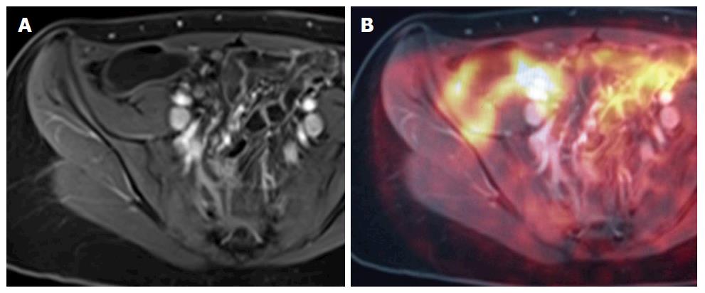
Figure 12 A and B demonstrate positron emission tomography magnetic resonance imaging.
The purpose of this figure is to demonstrate how an magnetic resonance image (A: Axial contrast enhanced T1 weighted image) and a PET image can be fused on a workstation to form an MR-PET image (B: Fused PET and MRI image) in cases where MR and PET images are acquired separately. MRI: Magnetic resonance imaging; PET: Positron emission tomography.
- Citation: Stanley E, Moriarty HK, Cronin CG. Advanced multimodality imaging of inflammatory bowel disease in 2015: An update. World J Radiol 2016; 8(6): 571-580
- URL: https://www.wjgnet.com/1949-8470/full/v8/i6/571.htm
- DOI: https://dx.doi.org/10.4329/wjr.v8.i6.571


