Copyright
©The Author(s) 2016.
World J Radiol. Jun 28, 2016; 8(6): 571-580
Published online Jun 28, 2016. doi: 10.4329/wjr.v8.i6.571
Published online Jun 28, 2016. doi: 10.4329/wjr.v8.i6.571
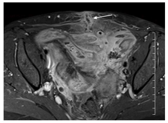
Figure 10 Axial contrast enhanced T1 weighted images demonstrate extensive inflammation and enhancement of small bowel and mesentery in the pelvis in a patient with acute Crohn’s disease.
In this image, there is good delineation of an enterocutaneous fistula extending from pelvic small bowel to the lower anterior abdominal wall in the midline (white arrow).
- Citation: Stanley E, Moriarty HK, Cronin CG. Advanced multimodality imaging of inflammatory bowel disease in 2015: An update. World J Radiol 2016; 8(6): 571-580
- URL: https://www.wjgnet.com/1949-8470/full/v8/i6/571.htm
- DOI: https://dx.doi.org/10.4329/wjr.v8.i6.571


