Copyright
©The Author(s) 2016.
World J Radiol. Jun 28, 2016; 8(6): 571-580
Published online Jun 28, 2016. doi: 10.4329/wjr.v8.i6.571
Published online Jun 28, 2016. doi: 10.4329/wjr.v8.i6.571
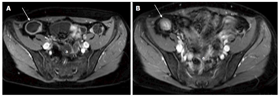
Figure 6 A and B: Axial contrast enhanced T1 weighted images of a 38-year-old female with Crohn’s disease.
A: shows a dilated terminal ileum with avid mural enhancement (white arrow) with further wall enhancing small bowel loops in the pelvis (a); B: Acquired following a course of Azithioprine treatment, and demonstrates a treatment response. The terminal ileum no longer demonstrates mural enhancement (white arrow). There is now relatively hyperintense T1 signal in the wall of the terminal ileum when compared with the mesenteric fat, consistent with fatty infiltration - the “Fat Halo sign”. Pelvic small bowel loops no longer display post contrast mural enhancemen (a).
- Citation: Stanley E, Moriarty HK, Cronin CG. Advanced multimodality imaging of inflammatory bowel disease in 2015: An update. World J Radiol 2016; 8(6): 571-580
- URL: https://www.wjgnet.com/1949-8470/full/v8/i6/571.htm
- DOI: https://dx.doi.org/10.4329/wjr.v8.i6.571


