Copyright
©The Author(s) 2016.
World J Radiol. Jun 28, 2016; 8(6): 571-580
Published online Jun 28, 2016. doi: 10.4329/wjr.v8.i6.571
Published online Jun 28, 2016. doi: 10.4329/wjr.v8.i6.571
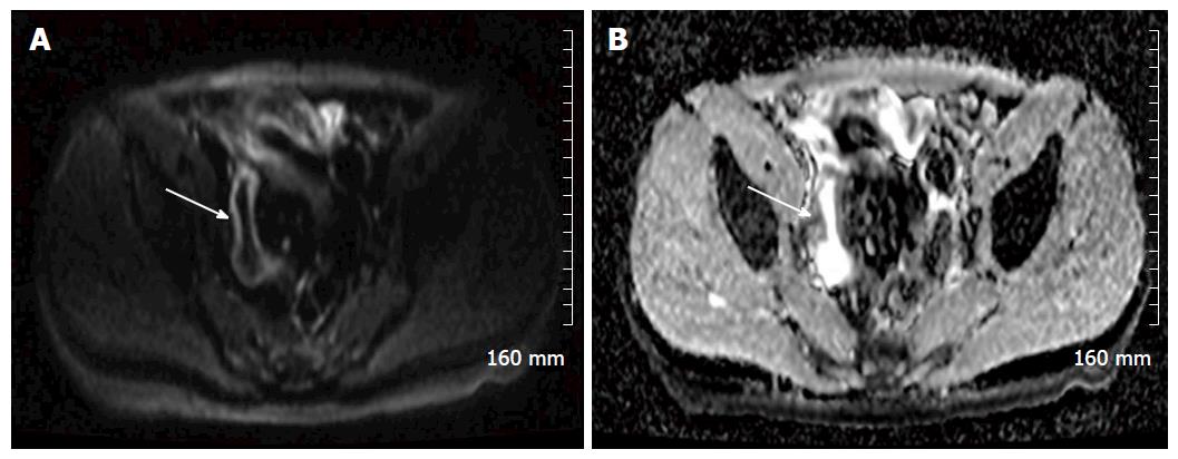
Figure 5 An example of restricted diffusion within the wall of an acutely inflamed distal ileum in a patient with acute Crohn’s disease.
A: A B 800 image, demonstrating hyperintense signal in the mucosa of the distal ileum (white arrow); B: The apparent diffusion coefficient map, and demonstrates corresponding hypointense signal (white arrow).
- Citation: Stanley E, Moriarty HK, Cronin CG. Advanced multimodality imaging of inflammatory bowel disease in 2015: An update. World J Radiol 2016; 8(6): 571-580
- URL: https://www.wjgnet.com/1949-8470/full/v8/i6/571.htm
- DOI: https://dx.doi.org/10.4329/wjr.v8.i6.571


