Copyright
©The Author(s) 2016.
World J Radiol. Jun 28, 2016; 8(6): 556-570
Published online Jun 28, 2016. doi: 10.4329/wjr.v8.i6.556
Published online Jun 28, 2016. doi: 10.4329/wjr.v8.i6.556
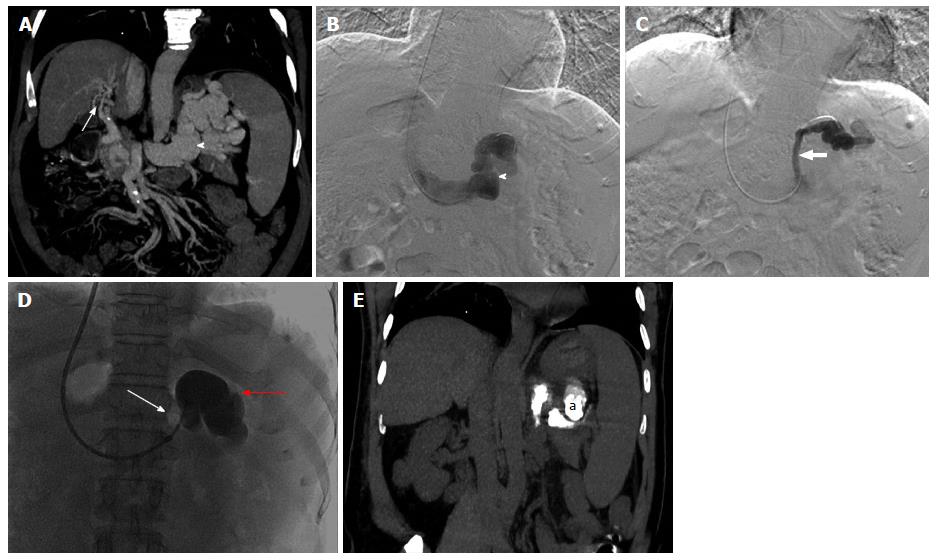
Figure 11 Fifty-year-old male patient, known case of extrahepatic portal vein obstruction with recurrent hepatic encephalopathy and hepatic parkinsonism.
Maximum intensity projection coronal post contrast CT image (A) and DSA images (B and C) showing portal cavernoma formation (arrow), a large gastorenal shunt (arrow head) and another small gastrorenal shunt identified within same patient. DSA image (D) showing balloon occluded (thin arrow) retrograde transvenous occlusion (BRTO) of only the large gastrorenal shunt (red arrow) through transjugular approach with the help of lipoidol-sclerosant foam. Post BRTO, non-contrast CT coronal image showing lipoidol cast (a) within the large gastrorenal shunt (E). BRTO: Balloon occluded trans-venous obliteration; CT: Computed tomography; DSA: Digital substraction angiography.
- Citation: Pargewar SS, Desai SN, Rajesh S, Singh VP, Arora A, Mukund A. Imaging and radiological interventions in extra-hepatic portal vein obstruction. World J Radiol 2016; 8(6): 556-570
- URL: https://www.wjgnet.com/1949-8470/full/v8/i6/556.htm
- DOI: https://dx.doi.org/10.4329/wjr.v8.i6.556


