Copyright
©The Author(s) 2016.
World J Radiol. Jun 28, 2016; 8(6): 556-570
Published online Jun 28, 2016. doi: 10.4329/wjr.v8.i6.556
Published online Jun 28, 2016. doi: 10.4329/wjr.v8.i6.556
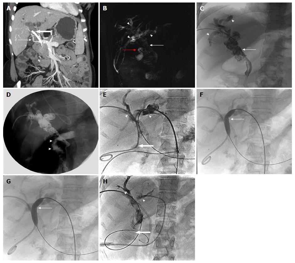
Figure 9 Twenty-five-years-old female, known case of extrahepatic portal vein obstruction with portal cavernoma cholangiopathy.
A: Maximum intensity projection post contrast coronal image showing portal cevernoma (arrow) and changes of PCC in the form of bilobar IHBRD with dilated CBD having stent in situ (block arrow); three-dimensional MRCP image (B) and PTC (C) showing dilated CBD with choledocholithiasis (thin arrow), IHBR dilatation with hepatolithiasis (arrow heads) and cholelithiasis (red arrow); D: Intraoperative cholangiogram after biliary diversion in the form of hepatico-jejunal anastomosis showing free flow of contrast into jejunal lumen (arrow heads); E: Follow-up PTC showing hepatico-jejunostomy anastomotic site stricture (block arrow) with upstream bilobar IHBR dilatation and hepatolithiasis (arrow heads) with faint contrast opacification of jejunal loop (block arrow); F and G: Fluoroscopic spot images showing progressive dilatation of anastomotic site stricture using conventional balloon (thin arrow) with assisted stone clearance; H: Post balloon dilatation and stone clearance, PTC showing relatively better contrast opacification of jejunal loop (block arrow) and relative reduction in amount of IHBRD and hepatolithiasis (arrow heads). CBD: Common bile duct; PCC: Portal cavernoma cholangiopathy; IHBRD: Intrahepatic biliary dilatation; MRCP: Magnetic resonance cholangiopancreatography; PTC: Percutaneous transhepatic cholangiogram.
- Citation: Pargewar SS, Desai SN, Rajesh S, Singh VP, Arora A, Mukund A. Imaging and radiological interventions in extra-hepatic portal vein obstruction. World J Radiol 2016; 8(6): 556-570
- URL: https://www.wjgnet.com/1949-8470/full/v8/i6/556.htm
- DOI: https://dx.doi.org/10.4329/wjr.v8.i6.556


