Copyright
©The Author(s) 2016.
World J Radiol. Jun 28, 2016; 8(6): 556-570
Published online Jun 28, 2016. doi: 10.4329/wjr.v8.i6.556
Published online Jun 28, 2016. doi: 10.4329/wjr.v8.i6.556
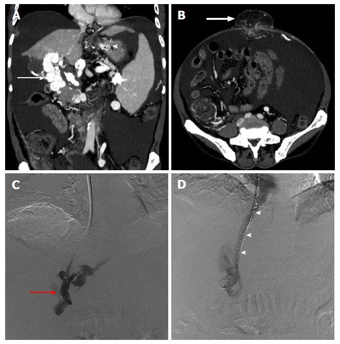
Figure 8 Sixty-year-old male patient, a known case of extrahepatic portal vein obstruction with history of recurrent upper gastrointestinal bleed and umbilical hernia, subjected to transjugular intrahepatic portosystemic shunt procedure in order to reduce blood loss during contemplated future surgical repair of umbilical hernia.
Maximum intensity projection coronal (A) and axial (B) post contrast CT images showing portal cavernoma (arrow), gastric varices (arrow head) and umbilical hernia (bold arrow). DSA images showing portal venogram (C) after accessing portal vein (red arrow) through transjugular approach and post TIPS stent deployment (arrow heads), diversion of portal flow into right atrium via hepatic vein (D). TIPS: Transjugular intra-hepatic portosystemic shunt; DSA: Digital substration angiography; CT: Computed tomography.
- Citation: Pargewar SS, Desai SN, Rajesh S, Singh VP, Arora A, Mukund A. Imaging and radiological interventions in extra-hepatic portal vein obstruction. World J Radiol 2016; 8(6): 556-570
- URL: https://www.wjgnet.com/1949-8470/full/v8/i6/556.htm
- DOI: https://dx.doi.org/10.4329/wjr.v8.i6.556


