Copyright
©The Author(s) 2016.
World J Radiol. Jun 28, 2016; 8(6): 556-570
Published online Jun 28, 2016. doi: 10.4329/wjr.v8.i6.556
Published online Jun 28, 2016. doi: 10.4329/wjr.v8.i6.556
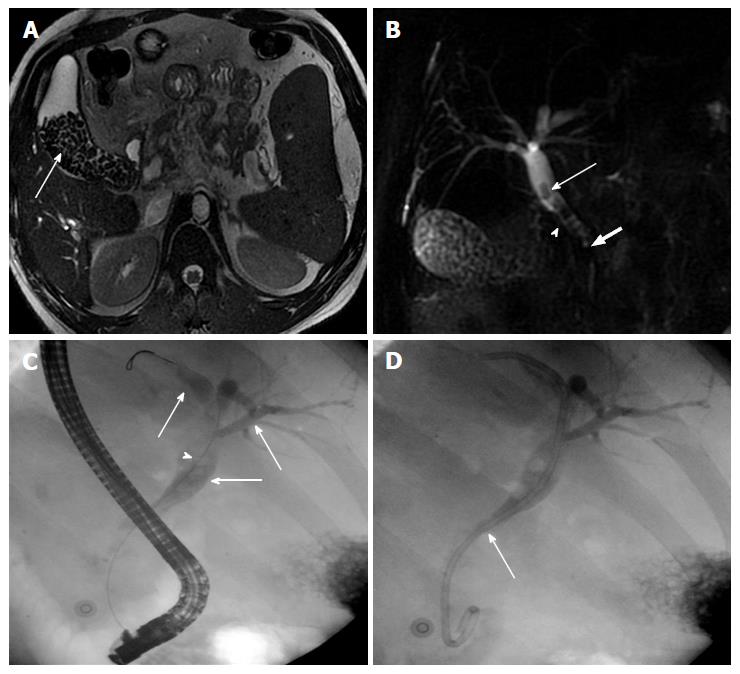
Figure 6 Cholelithiasis and choledocholithiasis.
A: MR Axial T2W gradient recalled echo image showing multiple variable sized calculi (arrow) filling the gallbladder; B: Thick-slab two-dimensional MRC image of the same patient depicting additionally choledocholithiasis (arrow). Also note the irregular CBD contour (arrow head) with angulated appearance of suprapancreatic CBD (block arrow) consistent with biliopathy; C: ERC of this patient shows dilated intra and extra-hepatic biliary system (thin arrow) with multiple filling defects (arrow heads) in CBD consistent with calculi; D: Post stone retrieval and biliary stent placement (thin arrow). MRC: Magnetic resonance cholangiography; CBD: Common bile duct; ERC: Endoscopic retrograde cholangiography.
- Citation: Pargewar SS, Desai SN, Rajesh S, Singh VP, Arora A, Mukund A. Imaging and radiological interventions in extra-hepatic portal vein obstruction. World J Radiol 2016; 8(6): 556-570
- URL: https://www.wjgnet.com/1949-8470/full/v8/i6/556.htm
- DOI: https://dx.doi.org/10.4329/wjr.v8.i6.556


