Copyright
©The Author(s) 2016.
World J Radiol. Jun 28, 2016; 8(6): 556-570
Published online Jun 28, 2016. doi: 10.4329/wjr.v8.i6.556
Published online Jun 28, 2016. doi: 10.4329/wjr.v8.i6.556
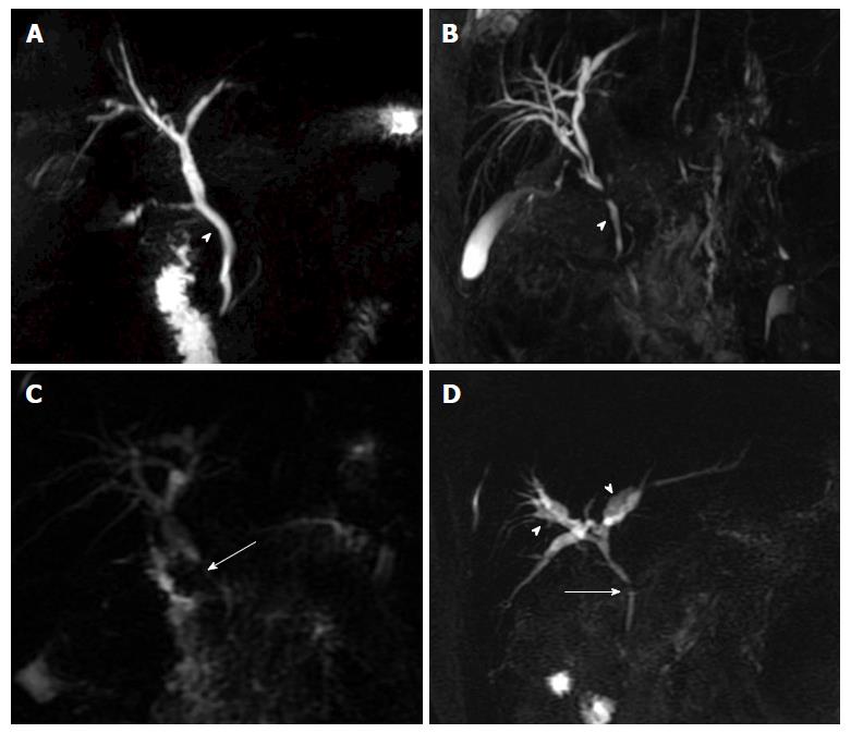
Figure 5 Portal biliopathy changes on magnetic resonance cholangiography.
Thick-slab two dimensional MRC images showing: A: Extrinsic nodular thumb-like impressions (arrow head) on CBD; B: Fine, wavy irregular contour (arrow head) of CBD; C: Stricture (arrow) in supra-pancreatic CBD with upstream dilatation; D: Angulated CBD (arrow) with smooth contour, patent primary confluence and intra-hepatic biliary ductal dilatation (arrow heads). MRC: Magnetic resonance cholangiography; CBD: Common bile duct.
- Citation: Pargewar SS, Desai SN, Rajesh S, Singh VP, Arora A, Mukund A. Imaging and radiological interventions in extra-hepatic portal vein obstruction. World J Radiol 2016; 8(6): 556-570
- URL: https://www.wjgnet.com/1949-8470/full/v8/i6/556.htm
- DOI: https://dx.doi.org/10.4329/wjr.v8.i6.556


