Copyright
©The Author(s) 2016.
World J Radiol. Jun 28, 2016; 8(6): 556-570
Published online Jun 28, 2016. doi: 10.4329/wjr.v8.i6.556
Published online Jun 28, 2016. doi: 10.4329/wjr.v8.i6.556
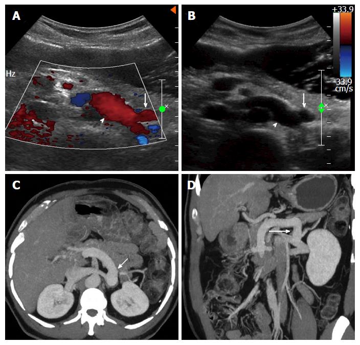
Figure 2 Proximal spleno-renal shunt.
Doppler (A) and gray-scale (B) ultrasound images showing a postoperative patient with patent proximal splenorenal shunt (arrow) draining into patent left renal vein (arrow head), axial (C) and coronal (D) thick maximum intensity projection images clearly demonstrating the shunt (arrow) patency.
- Citation: Pargewar SS, Desai SN, Rajesh S, Singh VP, Arora A, Mukund A. Imaging and radiological interventions in extra-hepatic portal vein obstruction. World J Radiol 2016; 8(6): 556-570
- URL: https://www.wjgnet.com/1949-8470/full/v8/i6/556.htm
- DOI: https://dx.doi.org/10.4329/wjr.v8.i6.556


