Copyright
©The Author(s) 2016.
World J Radiol. Jun 28, 2016; 8(6): 556-570
Published online Jun 28, 2016. doi: 10.4329/wjr.v8.i6.556
Published online Jun 28, 2016. doi: 10.4329/wjr.v8.i6.556
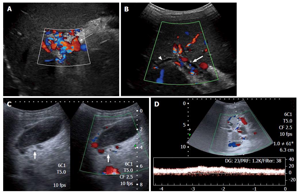
Figure 1 Ultrasonography and Doppler in extrahepatic portal vein obstruction.
A: Doppler showing multiple tortuous portoportal collaterals forming a portal cavernoma (arrow) and replacing the portal vein; B: USG with Doppler showing parabiliary collaterals surrounding the prominent CBD (block arrow) and intrahepatic biliary radicles (arrow head); C: Pericholecystic varices (block arrow) are also noted; D: Spectral Doppler reveals hepatopetal flow with monophasic waveform. USG: Ultrasonography; CBD: Common bile duct.
- Citation: Pargewar SS, Desai SN, Rajesh S, Singh VP, Arora A, Mukund A. Imaging and radiological interventions in extra-hepatic portal vein obstruction. World J Radiol 2016; 8(6): 556-570
- URL: https://www.wjgnet.com/1949-8470/full/v8/i6/556.htm
- DOI: https://dx.doi.org/10.4329/wjr.v8.i6.556


