Copyright
©The Author(s) 2016.
World J Radiol. Jun 28, 2016; 8(6): 537-555
Published online Jun 28, 2016. doi: 10.4329/wjr.v8.i6.537
Published online Jun 28, 2016. doi: 10.4329/wjr.v8.i6.537
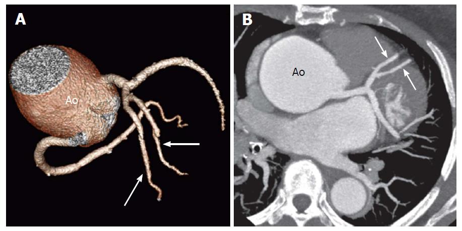
Figure 16 Duplication of the left anterior descending artery in a 52-year-old woman at preoperative evaluation[14].
Volume- rendered image (A) and oblique axial computed tomography image (B) show two equally sized LAD arteries (arrows). The Ao is noted to be aneurysmal. LAD: Left anterior descending; Ao: Aorta.
- Citation: Villa AD, Sammut E, Nair A, Rajani R, Bonamini R, Chiribiri A. Coronary artery anomalies overview: The normal and the abnormal. World J Radiol 2016; 8(6): 537-555
- URL: https://www.wjgnet.com/1949-8470/full/v8/i6/537.htm
- DOI: https://dx.doi.org/10.4329/wjr.v8.i6.537


