Copyright
©The Author(s) 2016.
World J Radiol. Jun 28, 2016; 8(6): 537-555
Published online Jun 28, 2016. doi: 10.4329/wjr.v8.i6.537
Published online Jun 28, 2016. doi: 10.4329/wjr.v8.i6.537
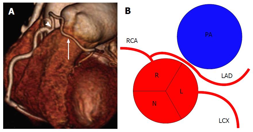
Figure 14 Left anterior desceding from right coronary artery[2].
A: Volume-rendered computed tomography image of LAD (arrow) artery arising from proximal RCA (arrowhead); B: Schematic representation. RCA: Right coronary artery; LCX: Left circumflex; LAD: Left anterior descending; R: Right sinus of Valsalva; L: Left sinus of Valsalva; N: Non-coronary sinus; PA: Pulmonary artery.
- Citation: Villa AD, Sammut E, Nair A, Rajani R, Bonamini R, Chiribiri A. Coronary artery anomalies overview: The normal and the abnormal. World J Radiol 2016; 8(6): 537-555
- URL: https://www.wjgnet.com/1949-8470/full/v8/i6/537.htm
- DOI: https://dx.doi.org/10.4329/wjr.v8.i6.537


