Copyright
©The Author(s) 2016.
World J Radiol. Jun 28, 2016; 8(6): 537-555
Published online Jun 28, 2016. doi: 10.4329/wjr.v8.i6.537
Published online Jun 28, 2016. doi: 10.4329/wjr.v8.i6.537
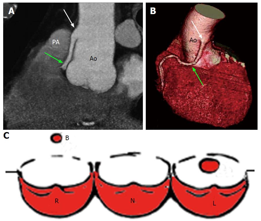
Figure 4 High take-off of right coronary artery in a 67-year-old man with equivocal exercise stress test[57].
Maximum intensity projection coronary CT angiogram (A) and three-dimensional volume-rendered CT reconstruction (B). Aortic step (white arrow) is seen to be the origin of RCA; RCA runs closely parallel to the proximal aorta (Ao) behind the pulmonary artery (PA). A: Regular position of left ostium; B: High position of right ostium; C: Schematic representation of high take-off. R: Right sinus of Valsalva; L: Left sinus of Valsalva; N: Non-coronary sinus; RCA: Right coronary artery; CT: Computed tomography.
- Citation: Villa AD, Sammut E, Nair A, Rajani R, Bonamini R, Chiribiri A. Coronary artery anomalies overview: The normal and the abnormal. World J Radiol 2016; 8(6): 537-555
- URL: https://www.wjgnet.com/1949-8470/full/v8/i6/537.htm
- DOI: https://dx.doi.org/10.4329/wjr.v8.i6.537


