Copyright
©The Author(s) 2016.
World J Radiol. May 28, 2016; 8(5): 530-536
Published online May 28, 2016. doi: 10.4329/wjr.v8.i5.530
Published online May 28, 2016. doi: 10.4329/wjr.v8.i5.530
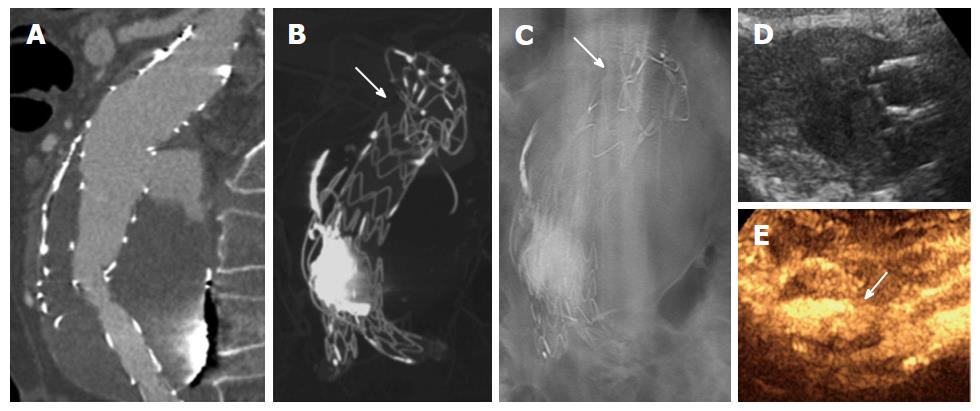
Figure 2 Summarization of the findings according to the different imaging techniques of a type III endoleak after graft dislocation.
(A) Computed tomography angiography with focal contrast-enhancement behind the graft main body during arterial phase contrast shows the presence of endoleak; computed tomography angiography multiplanar MIP reconstruction (B) and antero-posterior digital tomosynthesis of the abdomen (C) shows better visualization of the graft failure with complete dislocation and angulation of the graft main body (white harrows); Ultrasound image of the enlarged aneurismal sac in an axial scan (D) and the visualization of the endoleak with a contrast-enhanced ultrasound scan (arrow indicates the endoleak flux) (E).
- Citation: Mazzei MA, Guerrini S, Mazzei FG, Cioffi Squitieri N, Notaro D, de Donato G, Galzerano G, Sacco P, Setacci F, Volterrani L, Setacci C. Follow-up of endovascular aortic aneurysm repair: Preliminary validation of digital tomosynthesis and contrast enhanced ultrasound in detection of medium- to long-term complications. World J Radiol 2016; 8(5): 530-536
- URL: https://www.wjgnet.com/1949-8470/full/v8/i5/530.htm
- DOI: https://dx.doi.org/10.4329/wjr.v8.i5.530


