Copyright
©The Author(s) 2016.
World J Radiol. May 28, 2016; 8(5): 518-529
Published online May 28, 2016. doi: 10.4329/wjr.v8.i5.518
Published online May 28, 2016. doi: 10.4329/wjr.v8.i5.518
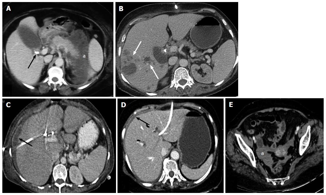
Figure 10 Complications.
A: Axial contrast enhanced CT scan showing bulky pancreas with peripancreatic necrotic collection (a) developing after PTBD in a patient of carcinoma of gall bladder (arrow); B: Axial contrast enhanced CT scan after PTBD showing multiple abscesses in right lobe of liver (arrows); C: Axial CT scan after PTBD showing perihepatic bilioma (arrow); D and E: Axial CT scans after PTBD showing hyperdense fluid in perihepatic (arrow) region and pelvis (a) suggesting hemoperitoneum. CT: Computed tomography; PTBD: Percutaneous transhepatic biliary drainage.
- Citation: Madhusudhan KS, Gamanagatti S, Srivastava DN, Gupta AK. Radiological interventions in malignant biliary obstruction. World J Radiol 2016; 8(5): 518-529
- URL: https://www.wjgnet.com/1949-8470/full/v8/i5/518.htm
- DOI: https://dx.doi.org/10.4329/wjr.v8.i5.518


