Copyright
©The Author(s) 2016.
World J Radiol. May 28, 2016; 8(5): 518-529
Published online May 28, 2016. doi: 10.4329/wjr.v8.i5.518
Published online May 28, 2016. doi: 10.4329/wjr.v8.i5.518
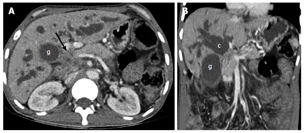
Figure 1 Axial (A) and coronal (B) contrast enhanced computed tomography scan of a patient with carcinoma of gall bladder showing mass (arrow) arising from the neck of gall bladder (g) involving common hepatic duct with patent primary biliary confluence (c).
- Citation: Madhusudhan KS, Gamanagatti S, Srivastava DN, Gupta AK. Radiological interventions in malignant biliary obstruction. World J Radiol 2016; 8(5): 518-529
- URL: https://www.wjgnet.com/1949-8470/full/v8/i5/518.htm
- DOI: https://dx.doi.org/10.4329/wjr.v8.i5.518


