Copyright
©The Author(s) 2016.
World J Radiol. May 28, 2016; 8(5): 513-517
Published online May 28, 2016. doi: 10.4329/wjr.v8.i5.513
Published online May 28, 2016. doi: 10.4329/wjr.v8.i5.513
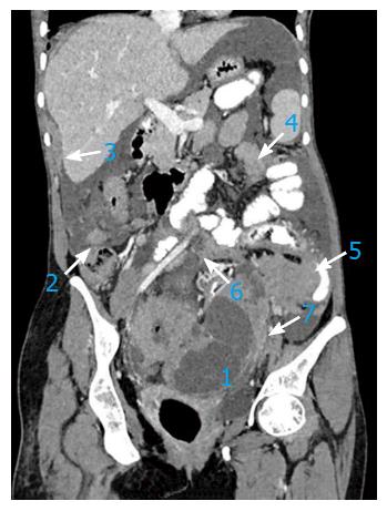
Figure 1 In a 67-year-old patient with advanced stage ovarian carcinoma, the post-contrast computed tomography coronal image depicts preferential spread of peritoneal implant involvement of rectouterine pouch (1), right paracolic gutter (2), right perihepatic space (3), inframesocolon (4), left paracolic gutter (5), mesentery (6) and mesosigmoid (7).
- Citation: Chandrashekhara SH, Triveni GS, Kumar R. Imaging of peritoneal deposits in ovarian cancer: A pictorial review. World J Radiol 2016; 8(5): 513-517
- URL: https://www.wjgnet.com/1949-8470/full/v8/i5/513.htm
- DOI: https://dx.doi.org/10.4329/wjr.v8.i5.513


