Copyright
©The Author(s) 2016.
World J Radiol. May 28, 2016; 8(5): 460-471
Published online May 28, 2016. doi: 10.4329/wjr.v8.i5.460
Published online May 28, 2016. doi: 10.4329/wjr.v8.i5.460
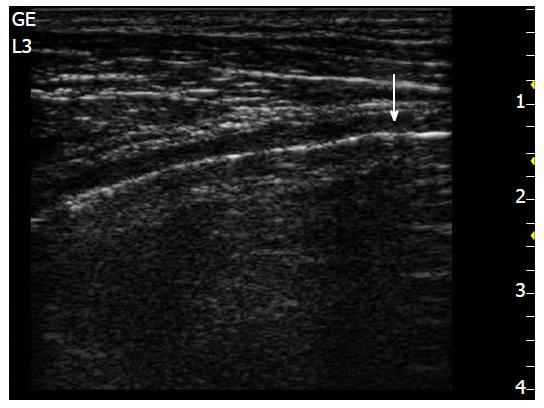
Figure 9 Lung point (arrow).
Is the precise area of the chest wall, where the regular reappearance of the lung sliding replaces the pneumothorax pattern and it corresponds to the point where the visceral and parietal pleura regain contact with each other (by image courtesy of Dr. Lucia Morelli).
- Citation: Liccardo B, Martone F, Trambaiolo P, Severino S, Cibinel GA, D’Andrea A. Incremental value of thoracic ultrasound in intensive care units: Indications, uses, and applications. World J Radiol 2016; 8(5): 460-471
- URL: https://www.wjgnet.com/1949-8470/full/v8/i5/460.htm
- DOI: https://dx.doi.org/10.4329/wjr.v8.i5.460


