Copyright
©The Author(s) 2016.
World J Radiol. May 28, 2016; 8(5): 434-448
Published online May 28, 2016. doi: 10.4329/wjr.v8.i5.434
Published online May 28, 2016. doi: 10.4329/wjr.v8.i5.434
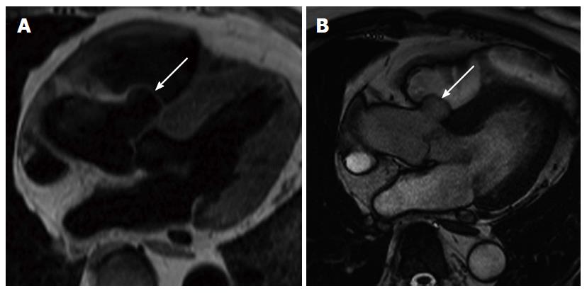
Figure 21 Sinus of Valsalva aneurysm.
A: Four chamber black blood magnetic resonance (MR) shows right sinus of Valsalva aneurysm (arrow) protruding into the right ventricular outflow tract (RVOT); B: Four chamber steady state free precession MR shows right sinus of Valsalva aneurysm (arrow) protruding into the RVOT.
- Citation: Kalisz K, Rajiah P. Radiological features of uncommon aneurysms of the cardiovascular system. World J Radiol 2016; 8(5): 434-448
- URL: https://www.wjgnet.com/1949-8470/full/v8/i5/434.htm
- DOI: https://dx.doi.org/10.4329/wjr.v8.i5.434


