Copyright
©The Author(s) 2016.
World J Radiol. May 28, 2016; 8(5): 434-448
Published online May 28, 2016. doi: 10.4329/wjr.v8.i5.434
Published online May 28, 2016. doi: 10.4329/wjr.v8.i5.434
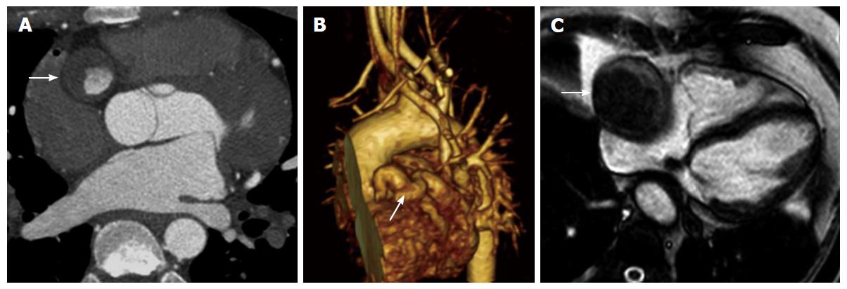
Figure 19 Coronary artery aneurysm.
A: Axial contrast enhanced computed tomography (CT) in a patient with a history of Kawasaki disease shows aneurysm of the right coronary artery (arrow) with significant mural thrombus; B: Three-dimensional volume rendered CT shows aneurysm of the right coronary artery (arrow); C: Four chamber magnetic resonance steady state free precession image in patient with saphenous vein graft to the posterior descending artery shows aneurysm of the graft (arrow) with mural thrombus and mass effect posteriorly onto the adjacent right atrium.
- Citation: Kalisz K, Rajiah P. Radiological features of uncommon aneurysms of the cardiovascular system. World J Radiol 2016; 8(5): 434-448
- URL: https://www.wjgnet.com/1949-8470/full/v8/i5/434.htm
- DOI: https://dx.doi.org/10.4329/wjr.v8.i5.434


