Copyright
©The Author(s) 2016.
World J Radiol. May 28, 2016; 8(5): 434-448
Published online May 28, 2016. doi: 10.4329/wjr.v8.i5.434
Published online May 28, 2016. doi: 10.4329/wjr.v8.i5.434
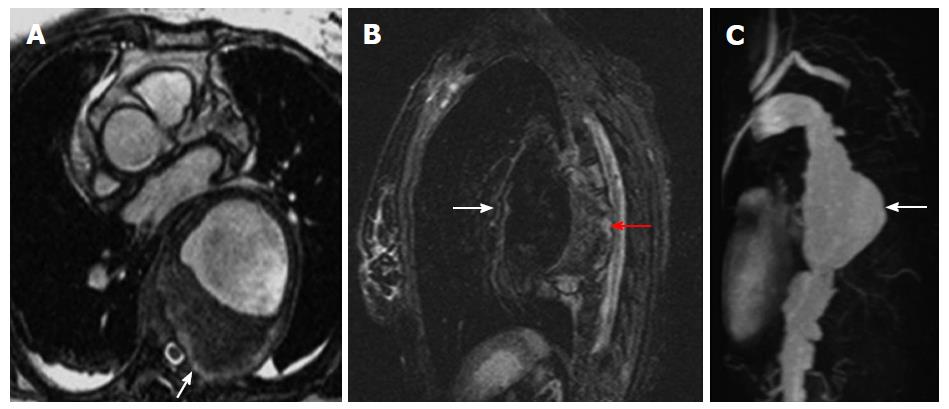
Figure 9 Mycotic aneurysm.
A: Axial contrast enhanced magnetic resonance angiography (MRA) shows large proximal descending aortic aneurysm with eccentric thrombus and invasion of adjacent thoracic vertebral body (arrow); B: Sagittal short tau inversion recovery shows large descending aortic aneurysm (white arrow) with high signal within the wall posteriorly (red arrow) indicating acute inflammation; C: Sagittal contrast enhanced MRA maximum intensity projection reconstruction shows proximal descending aortic mycotic aneurysm (arrow) extending posteriorly.
- Citation: Kalisz K, Rajiah P. Radiological features of uncommon aneurysms of the cardiovascular system. World J Radiol 2016; 8(5): 434-448
- URL: https://www.wjgnet.com/1949-8470/full/v8/i5/434.htm
- DOI: https://dx.doi.org/10.4329/wjr.v8.i5.434


