Copyright
©The Author(s) 2016.
World J Radiol. May 28, 2016; 8(5): 434-448
Published online May 28, 2016. doi: 10.4329/wjr.v8.i5.434
Published online May 28, 2016. doi: 10.4329/wjr.v8.i5.434
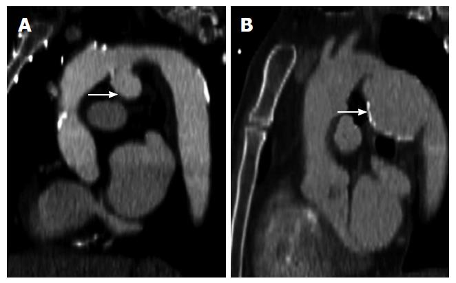
Figure 8 Ductus aneurysm.
A: Sagittal contrast enhanced computed tomography (CT) shows pseudoaneurysm (arrow) arising from the aortic isthmus and projecting inferiorly in a patient with a history of remote thoracic trauma; B: Sagittal contrast enhanced CT shows large, peripherally calcified ductus aneurysm (arrow).
- Citation: Kalisz K, Rajiah P. Radiological features of uncommon aneurysms of the cardiovascular system. World J Radiol 2016; 8(5): 434-448
- URL: https://www.wjgnet.com/1949-8470/full/v8/i5/434.htm
- DOI: https://dx.doi.org/10.4329/wjr.v8.i5.434


