Copyright
©The Author(s) 2016.
World J Radiol. May 28, 2016; 8(5): 434-448
Published online May 28, 2016. doi: 10.4329/wjr.v8.i5.434
Published online May 28, 2016. doi: 10.4329/wjr.v8.i5.434
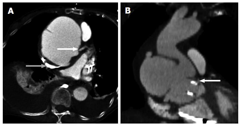
Figure 2 Giant aortic aneurysm with vascular compression.
A: Axial contrast enhanced computed tomography (CT) shows giant aortic aneurysm with dissection resulting in compression of the left main coronary artery (LMCA) ostium (white arrow) and superior vena cava (gray arrow); B: Coronal contrast enhanced CT shows giant aneurysm with dissection compressing the LMCA ostium (white arrow).
- Citation: Kalisz K, Rajiah P. Radiological features of uncommon aneurysms of the cardiovascular system. World J Radiol 2016; 8(5): 434-448
- URL: https://www.wjgnet.com/1949-8470/full/v8/i5/434.htm
- DOI: https://dx.doi.org/10.4329/wjr.v8.i5.434


