Copyright
©The Author(s) 2016.
World J Radiol. Apr 28, 2016; 8(4): 410-418
Published online Apr 28, 2016. doi: 10.4329/wjr.v8.i4.410
Published online Apr 28, 2016. doi: 10.4329/wjr.v8.i4.410
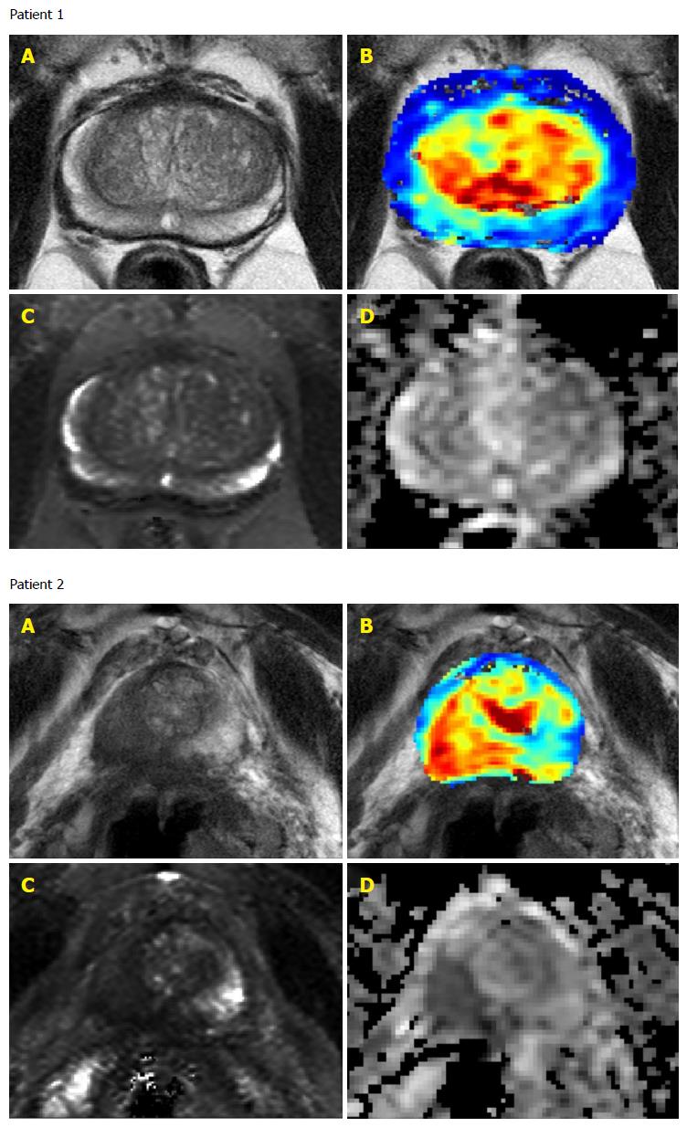
Figure 1 Baseline multiparametric magnetic resonance imaging images of two patients with Gleason score 6 low-risk prostate cancer but with different index of suspicion from multiparametric magnetic resonance imaging.
Patient 1 had an IOS of 1 on their baseline MRI scan and Patient 2 had an IOS of 5 on their baseline MRI scan. The mpMRI images shown for each patient are: A: T2-weighted image; B: DCE-parameter overlay (Amplitude); C: Quantitative T2 map; D: ADC map. mpMRI: Multiparametric magnetic resonance imaging; MRI: Magnetic resonance imaging; IOS: Index of suspicion; ADC: Apparent diffusion coefficient; DCE: Dynamic contrast-enhanced.
- Citation: Vos LJ, Janoski M, Wachowicz K, Yahya A, Boychak O, Amanie J, Pervez N, Parliament MB, Pituskin E, Fallone BG, Usmani N. Role of serial multiparametric magnetic resonance imaging in prostate cancer active surveillance. World J Radiol 2016; 8(4): 410-418
- URL: https://www.wjgnet.com/1949-8470/full/v8/i4/410.htm
- DOI: https://dx.doi.org/10.4329/wjr.v8.i4.410


