Copyright
©The Author(s) 2016.
World J Radiol. Apr 28, 2016; 8(4): 378-389
Published online Apr 28, 2016. doi: 10.4329/wjr.v8.i4.378
Published online Apr 28, 2016. doi: 10.4329/wjr.v8.i4.378
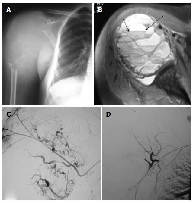
Figure 4 Preoperative embolisation of aneurysmal bone cyst of humerus.
Antero-posterior (A) plain radiograph of right shoulder show expansile lytic destructive lesion involving proximal end ofright humerus, suggestive of aneurysmal bone cyst; Axial (B) T2-weighted fat suppressed magnetic resonance image showlesion is hyperintense with fluid-fluid levels within it (arrow); Digital substraction angiography (C) image of right circumflex humeral artery show multiple feeders supplying the tumor with marked tumor blush; Post embolisation angiogram (D) of circumflex humeral artery show near complete reduction of tumor blush.
- Citation: Jha R, Sharma R, Rastogi S, Khan SA, Jayaswal A, Gamanagatti S. Preoperative embolization of primary bone tumors: A case control study. World J Radiol 2016; 8(4): 378-389
- URL: https://www.wjgnet.com/1949-8470/full/v8/i4/378.htm
- DOI: https://dx.doi.org/10.4329/wjr.v8.i4.378


