Copyright
©The Author(s) 2016.
World J Radiol. Apr 28, 2016; 8(4): 378-389
Published online Apr 28, 2016. doi: 10.4329/wjr.v8.i4.378
Published online Apr 28, 2016. doi: 10.4329/wjr.v8.i4.378
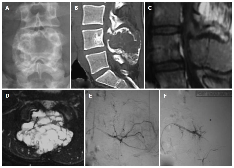
Figure 3 Preopertaive embolisation of spinal aneurysmal bone cyst.
Antero-posterior (A) plain radiograph of lumbo-sacral spine show expansile lytic lesion involving the posterior elements of L4 vertebra suggestive of aneurysmal bone cyst; Sagittal (B) 3D-reformatted non-contrast computed tomography of lumbo-sacral spine show lytic expansile destructive lesion involving posterior elements of L4 vertebra; Sagittal (C) and axial (D) T2-weighted fat saturated magnetic resonance images show T2 hyperintense lesion involving posterior element of L4 vertebra with blood-fluid levels (arrow); Pre embolisation non-selective digital substraction angiography (DSA) (E) image of L3 lumbar artery showmultiple feeders supplying the tumor with tumor blush; Post embolisation DSA (F) image show more than 90% reduction in tumor blush (asterix).
- Citation: Jha R, Sharma R, Rastogi S, Khan SA, Jayaswal A, Gamanagatti S. Preoperative embolization of primary bone tumors: A case control study. World J Radiol 2016; 8(4): 378-389
- URL: https://www.wjgnet.com/1949-8470/full/v8/i4/378.htm
- DOI: https://dx.doi.org/10.4329/wjr.v8.i4.378


