Copyright
©The Author(s) 2016.
World J Radiol. Apr 28, 2016; 8(4): 342-354
Published online Apr 28, 2016. doi: 10.4329/wjr.v8.i4.342
Published online Apr 28, 2016. doi: 10.4329/wjr.v8.i4.342
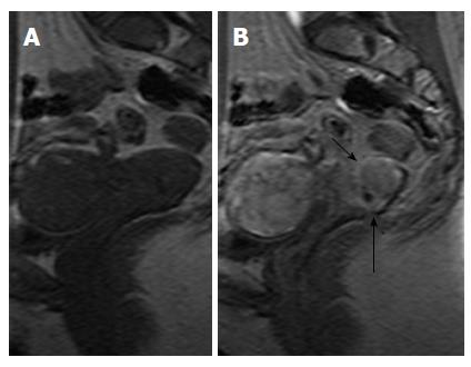
Figure 11 Dynamic contrast enhanced images, cervical cancer.
Sagittal pre- (A) and early arterial (30 s) post contrast (B) images. Cervical tumor borders are clearly delineated on the enhanced image (arrows in B). Note early arterial enhancement of cervical tumor in (B) synchronous with that of myometrium.
- Citation: Bourgioti C, Chatoupis K, Moulopoulos LA. Current imaging strategies for the evaluation of uterine cervical cancer. World J Radiol 2016; 8(4): 342-354
- URL: https://www.wjgnet.com/1949-8470/full/v8/i4/342.htm
- DOI: https://dx.doi.org/10.4329/wjr.v8.i4.342


