Copyright
©The Author(s) 2016.
World J Radiol. Apr 28, 2016; 8(4): 342-354
Published online Apr 28, 2016. doi: 10.4329/wjr.v8.i4.342
Published online Apr 28, 2016. doi: 10.4329/wjr.v8.i4.342
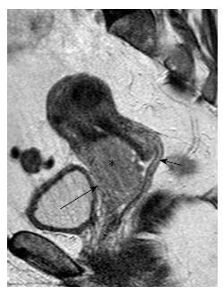
Figure 2 Cervical cancer, stage IIA.
Sagittal T2-weighted image shows a large cervical mass (asterisk) invading the anterior vaginal wall (long arrow). Preservation of the low signal intensity of the posterior vaginal fornix (small arrow) excludes invasion of posterior wall.
- Citation: Bourgioti C, Chatoupis K, Moulopoulos LA. Current imaging strategies for the evaluation of uterine cervical cancer. World J Radiol 2016; 8(4): 342-354
- URL: https://www.wjgnet.com/1949-8470/full/v8/i4/342.htm
- DOI: https://dx.doi.org/10.4329/wjr.v8.i4.342


