Copyright
©The Author(s) 2016.
World J Radiol. Mar 28, 2016; 8(3): 322-330
Published online Mar 28, 2016. doi: 10.4329/wjr.v8.i3.322
Published online Mar 28, 2016. doi: 10.4329/wjr.v8.i3.322
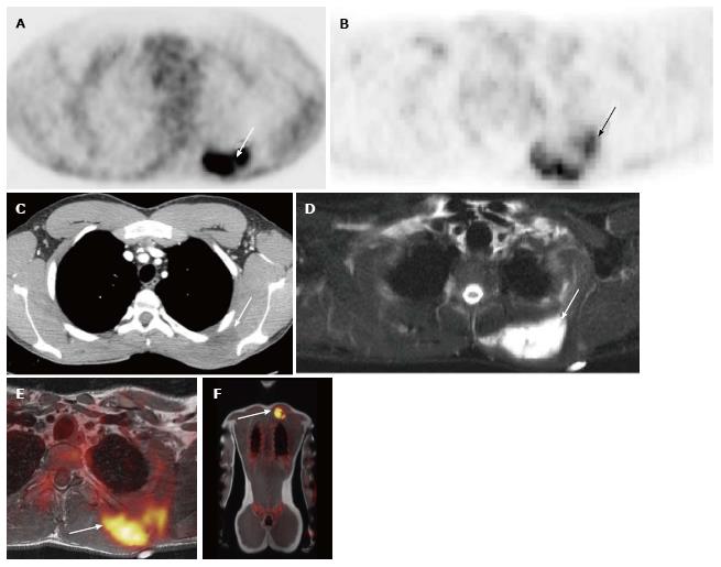
Figure 1 19-year-old male with undifferentiated malignant small round cell sarcoma.
Axial image from the PET-CT examination (A) shows intense uptake in mass in the left upper back (white arrow); axial image from the PET-MRI examination (B) shows similar FDG uptake in this region (white arrow); axial CT image from the PET-CT (C) shows a slightly hypodense mass in this region (white arrow). This mass is much better seen on the axial T2 fat suppressed image from the PET-MRI (D) (white arrow). Fused PET-MRI images [T1-weighted axial (E) and whole body coronal (F) MR sequences] again show intense FDG uptake associated with the mass in the left upper back (white arrows). PET-MRI: Positron emission tomography-magnetic resonance imaging; CT: Computed tomography; FDG: Fluorodeoxyglucose.
- Citation: Pugmire BS, Guimaraes AR, Lim R, Friedmann AM, Huang M, Ebb D, Weinstein H, Catalano OA, Mahmood U, Catana C, Gee MS. Simultaneous whole body 18F-fluorodeoxyglucose positron emission tomography magnetic resonance imaging for evaluation of pediatric cancer: Preliminary experience and comparison with 18F-fluorodeoxyglucose positron emission tomography computed tomography. World J Radiol 2016; 8(3): 322-330
- URL: https://www.wjgnet.com/1949-8470/full/v8/i3/322.htm
- DOI: https://dx.doi.org/10.4329/wjr.v8.i3.322


