Copyright
©The Author(s) 2016.
World J Radiol. Mar 28, 2016; 8(3): 316-321
Published online Mar 28, 2016. doi: 10.4329/wjr.v8.i3.316
Published online Mar 28, 2016. doi: 10.4329/wjr.v8.i3.316
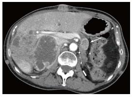
Figure 1 Fifty-four-year-old woman with pancreatic metastasis from retroperitoneal leiomyosarcoma.
Axial contrast-enhanced computed tomography image of the upper abdomen in the arterial phase demonstrates a hypovascular metastasis with central necrosis in the pancreatic body (arrow). Note the hepatic, right adrenal and subcutaneous metastasis.
- Citation: Suh CH, Keraliya A, Shinagare AB, Kim KW, Ramaiya NH, Tirumani SH. Multidetector computed tomography features of pancreatic metastases from leiomyosarcoma: Experience at a tertiary cancer center. World J Radiol 2016; 8(3): 316-321
- URL: https://www.wjgnet.com/1949-8470/full/v8/i3/316.htm
- DOI: https://dx.doi.org/10.4329/wjr.v8.i3.316


