Copyright
©The Author(s) 2016.
World J Radiol. Mar 28, 2016; 8(3): 298-307
Published online Mar 28, 2016. doi: 10.4329/wjr.v8.i3.298
Published online Mar 28, 2016. doi: 10.4329/wjr.v8.i3.298
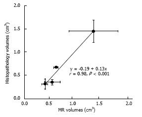
Figure 7 A close correlation was found between in situ magnetic resonance imaging and postmortem renal ablation volumes.
The bars in the x- and y-axes represent the standard error of the means. The ablated volumes after single sonication at 3000J and 4400J were not significantly different, while volumes at double sonications were significantly larger on both 4400J than 3000J. Furthermore, the effect of double sonications at 4400J was significantly larger than the other energies.
- Citation: Saeed M, Krug R, Do L, Hetts SW, Wilson MW. Renal ablation using magnetic resonance-guided high intensity focused ultrasound: Magnetic resonance imaging and histopathology assessment. World J Radiol 2016; 8(3): 298-307
- URL: https://www.wjgnet.com/1949-8470/full/v8/i3/298.htm
- DOI: https://dx.doi.org/10.4329/wjr.v8.i3.298


